

Inhibition of the latter system can therefore also cause dilation. Whereas stimulation of the parasympathetic system, known for "rest and digest" functions, causes constriction. Stimulation of the autonomic nervous system's sympathetic branch, known for triggering "fight or flight" responses when the body is under stress, induces pupil dilation. The iris is made of two types of muscle: a ring of sphincter muscles that encircle and constrict the pupil down to a couple of millimeters across to prevent too much light from entering and a set of dilator muscles laid out like bicycle spokes that can expand the pupil up to eight millimeters-approximately the diameter of a chickpea-in low light. Specifically, it dictates the movement of the iris to regulate the amount of light that enters the eye, similar to a camera aperture. But a different, older part of the nervous system-the autonomic-manages the continuous tuning of pupil size (along with other involuntary functions such as heart rate and perspiration).

The visual cortex in the back of the brain assembles the actual images we see. He views the dilations as a by-product of the nervous system processing important information. "Nobody really knows for sure what these changes do," says Stuart Steinhauer, director of the Biometrics Research Lab at the University of Pittsburgh School of Medicine. And they do this without knowing exactly why our eyes behave this way. In fact, pupil dilation correlates with arousal so consistently that researchers use pupil size, or pupillometry, to investigate a wide range of psychological phenomena. They also betray mental and emotional commotion. After one hour there was dilation of the constricted pupil and improvement in the ptosis.What do an orgasm, a multiplication problem and a photo of a dead body have in common? Each induces a slight, irrepressible expansion of the pupils in our eyes.įor more than a century scientists have known that our eyes' pupils respond to more than changes in light. One drop of apraclonidine 1% was applied to both eyes.
#POSSIBLE CAUSES UNEQUAL PUPIL SIZE FULL#
Ocular motility was full and funduscopy showed healthy optic discs. Visual acuity was 6/9 in both eyes and intraocular pressures were 12 mm Hg and 14 mm Hg in the right and left, respectively. Both pupils constricted to light and accommodation and there was no relative afferent pupillary defect. On examination she had a partial right upper lid ptosis (fig 1) and her right pupil, which was the abnormal one, was constricted and showed a dilatation lag.

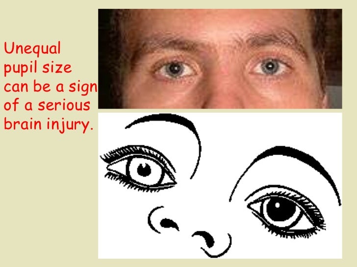
She denied any history of trauma, neck pain, weight loss, diplopia, or anhydrosis. She had visited her primary care doctor because of an upper respiratory tract infection and was unconcerned about her unequal pupil size (anisocoria), stating that she “has had odd pupils and a droopy right lid since being a teenager.” She had no ocular or medical history and was not taking any oral or topical medication.


 0 kommentar(er)
